Well, as any orthopedist will tell you, clinic is a necessary evil to spend so you can spend time in the OR. After several days of clinic, I was chomping at the bit to get into the action.
Disclaimer: The purpose of this blog is to share my experiences and perhaps help a future volunteer prepare for the realities of the medical environment outside the confines of home. I remain impressed – perhaps even in awe of the work and professionalism of my hosts. If any part of this story does not reflect that, it is a problem of my command of the English language, not their efforts!
The day began with a typical walk to the hospital. I was expecting “conference” which sounds like multidisciplinary grand rounds, but alas, it had been canceled. I walked up for resident rounds, but it was not scheduled for Fridays. I was, needless to say, a bit frustrated that after several days I still found myself struggling with understanding the weekly schedule. I think the miscommunications are largely reflective on the holiday schedule and current skeleton crew. I managed to locate a resident who felt there would be need for my help in the OR today.
9:00 came and went. 10:00 arrived to little action. It is quite a change for a type A personality surgeon to sit and wait. I am used to my 7:30 start meaning we are cutting before or by 7:30. I channeled my inner island time and waited.
I met a year 4 resident – who actually had been in clinic the day before. He discussed the OR schedule and seemed pleased I was available.
Today’s case list:
1-Closed distal femur fracture – 7 days old
2-Closed thumb dislocation – 2 days old (canceled yesterday)
3-Closed both bone forearm fracture – 5 days old
4-Closed bimalleolar ankle fracture – transferred from outside hospital, 2 days old.
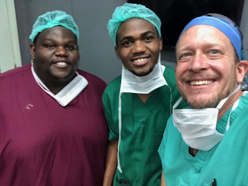
We went into the room to start the distal femur. To my surprise (pleasant), it was just me and resident. I tried to follow his lead – nothing is quite the same as the States. We donned a plastic apron over our scrubs – this is due to the cloth gowns, which while sterile are not water repellent. We had a “timeout” at bedside and introductions while the patient was awake. He then underwent spinal anesthesia. We applied a non sterile tourniquet (same as at home) and went to scrub. Interestingly, no trays, packs, etc were yet open.
After a scrub at the sink with detergent and water, we returned to the OR. There, we each received a handful of alcohol to finish our scrub. The back table now had a basic drape set, and 3 gowns opened. We each dried our hands, and grabbed a gown. After donning a gown, we each put on our own gloves (different from the US where a scrub tech is there to ensure I don’t screw up the sterile field). Now, the resident prepped the leg with an alcohol scrub and betadine paint.
Now to draping. The drape packs currently available are laparotomy packs. They are functional but lack the ease of use of extremity packs back home. My resident led me through draping while I added some pointer due to the case at hand. We inflated the tourniquet and were off and running.
[Sorry to be technical for the non medical…]
We made a standard lateral approach to the distal femur. Pre-op xrays suggested intra-articular involvement and multiple metaphyseal fragments. Pre-op positioning was less than ideal for these xrays, but due to cost concerns repeat films and CT are not an option.
Needless to say, the pre-operative xrays did not fully delineate the explosion we found at the distal metaphysis. The patella was comminuted but well-aligned. The condyles were split and the medial condyle was entrapped in soft tissue. My teaching points focused on blood flow to the bone and soft tissue preservation – techniques that are hard to follow when operating under somewhat challenging conditions. But we persevered.
We reconstructed the joint surface but struggled mightily with the metaphyseal reduction. Finally, we found missing fragments of bone entrapped in the quadriceps. This proved the missing link, and we were able to reconstruct the anterior femur allowing us to re-establish length and rotation. Length, angle, rotation – my mantra.
At this point dear friends, you are breathing a sigh of relief. But the true challenges are beginning. The scarcity of instrumentation and hardware is the true point of improvisation and challenge. We sort through plates and screws to piece together an appropriate construct. We have boxes of various plates and miscellaneous screws. Everything is reused – maximizing the availability of implants but creating a organizational challenge.
At last, the femur is fixed with plates and screws. Fist bumps for all (a personal post-fiction celebration of mine – shared with many of you) and the case is completed.
The next two cases are canceled due to financial concerns. It is approaching 2:30.
Next up – the ankle fracture. Armed with the experience of scrubbing a case, I take the lead in my wheel house – the foot and ankle. The resident is surprisingly timid for this case, and I learn that this case is not as common, as many ankles are treated closed with casting.
We face a high comminuted Weber C, syndesmosis disruption, bimalleolar ankle. I show him the approach, discuss AO techniques, and of course soft tissue preservation. The reduction at the fibula is somewhat difficult, but we obtain excellent alignment.
The implant issues begin again. We find a suitable plate, but no screws under 24 cm can be found. Not to be defeated, the bolt cutters are found, and we “trim” the longer screws to fit our needs. The fibula looks great. Similarly, the medial side and syndesmosis are fixed. I demonstrate closure techniques and the case concludes. Fist bumps – and more importantly, a successful surgery – and happy resident.
Which brings us to case 3. During case 2, the on call resident took a femur/tibia fracture to the OR down the hall. It had been transferred from an outside hospital and was over 24 hours old. The patient had a large laceration to the posterior knee that was sutured at the outside location and a last hemoglobin of 7 (done last night). He was pale, the leg was markedly swollen, and no distal pulses were palpable.
The resident (year 2) was rightfully very concerned and asked me to take a look. He had likely compartment syndrome of the thigh and leg as well as no capillary refill, decreased sensation below the knee, and a large hematoma around the laceration site. Very very bad.
Unfortunately, our fears were realized. We explored the popliteal fossa and found a huge hematoma. The resident calmly isolated the neurovascular bundle and it was not obviously damaged, but no pulse could be found. My best guess is the patient had a massive injury, significant bleeding, and developed compartment syndrome sometime yesterday. The distal musculature was dead. The artery below the knee was likely thrombosed. The sense of dread was palpable…
We spoke with staff, but due to consent concerns, could not proceed with amputation. Necrotic muscle was excised and the skin actually had some viability. An ex-fix to stabilize the leg was applied, but I fear this story has a poor ending. Hopefully, we can get consent and proceed with amputation before infection sets in. I hold my breath.
That concludes an eventful, difficult, triumphant, defeated, creative, frustrating, elating, and full day. I am out of adjectives – and it was just three cases. My walk home was definitely introspective. The demand outweighs the supply. Does this trip, this time, even my cut screws matter?
I conclude with a quote (or misquote) from Mother Teresa that adds warmth to the day and definitely gives me power to head back in Monday full of vigor, spit, salt and sugar!
“We ourselves feel that what we are doing is just a drop in the ocean. But the ocean would be less because of that missing drop.”
Until next week, keep dripping friends!